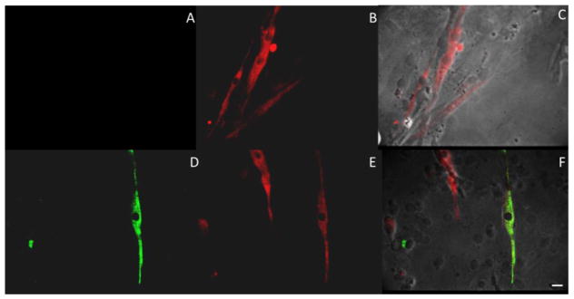
Figure 4.
Immunostaining of 12 DIV myotubes for adult MyHC isoforms. Panels (A and D) show staining for adult MyHC type I isoform for two separate coverslips. Panels (B and E) show staining for adult MyHC type IIb isoforms for those same myotubes. Panels (C and F) are the composite images of those cultures. Cultures indicate at 12 DIV 73.89 ± 4.6% of myotubes stain for type IIb and approximately 10.67 ± 1.1% of those also stain for type I isoform. Scale Bar 20 μm.

