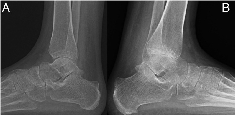
Figure 1.

Plain radiographs of the ankle 6 months apart. (A) Demonstrating early changes; however, the joint spaces are still relatively well preserved. There is dramatic progression in a span of 6 months as seen in (B) showing complete loss of joint space in the posterior subtalar articular surfaces with subchondral cysts and sclerosis.
