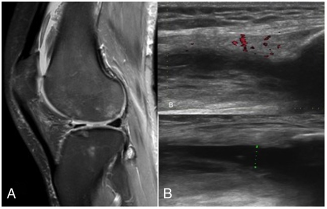
Figure 8.

Unenhanced non-arthrographic MRI of the knee showing a moderate knee joint effusion with ongoing active synovitis. Multifocal areas of chondral damage are appreciated (A). An ultrasound scan of the same knee demonstrating a significant knee joint effusion with suprapatellar extension and evidence of florid ongoing synovitis, as seen in (B) with increased flow on Doppler assessment. A diagnostic aspirate to exclude septic arthritis was performed at this stage (B and C).
