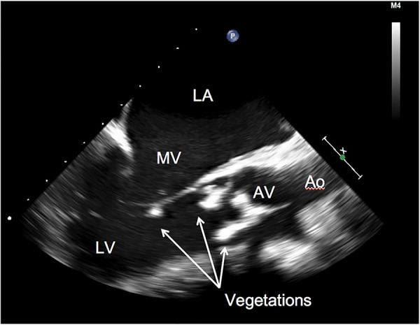
Figure 2.

Transoesophageal echocardiogram before surgical intervention. There are two elongated, echodense masses on the ventricular side of the aortic valve. Softer appearing density on the tip of the posterior leaflet of the mitral valve was also seen (arrows). Ao, ascending aorta; AV, aortic valve; LA, left atrium; LV, left ventricle; MV, mitral valve.
