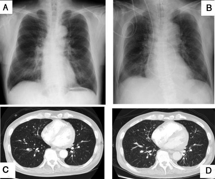
Figure 1.
Imaging findings of the chest. (A) Chest X-ray on standing at admission. (B) Last chest X-ray taken in supine position ∼5 hours prior to death. (C) Lung fields were grossly normal on CT scan at admission. (D) CT scan on day 11 revealed diffuse minute granular shadows in all lung fields and increased right ventricle volume.

