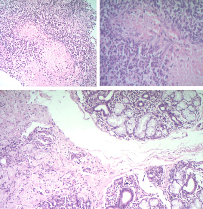
Figure 5.
Histological sections reveal islands of salivary gland tissue with intralobular and extralobular lymphocytic infiltrate. Areas of extensive oedema around the squamous epithelium with neutrophilic infiltration. Submucosa revealing medium arteriole vasculitis, obliteration of lumen with necrosis.

