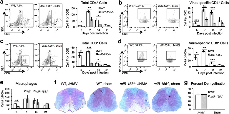
Fig. 2.

JHMV-infected miR-155 −/− mice demonstrated reduced CNS T cell infiltration. WT and miR-155 −/− mice were infected i.c. with JHMV (200 PFU). Mice from each group were sacrificed 5, 7, 14, and 21 days p.i., and brains were collected. Flow analysis indicated reduced infiltration of total CD4+ T cells (a) and CD8+ T cells (c), as well as reduced virus-specific CD4+ T cells (b) and CD8+ T cells (d), as determined by tetramer staining [95, 96]. In contrast, while macrophage (CD45 + F4/80hi) infiltration into the CNS was lower in miR-155 −/− mice at 5 days p.i. (e), the levels were similar at later time points. Representative spinal cords from JHMV-infected and sham-infected mice stained with LFB at day 14 p.i. showed similar levels of demyelination between infected WT mice (35.1 + 4.9 %, n = 4) and miR-155 −/− mice (36.7 + 4.3, n = 4) whereas no demyelination is observed in sham-infected animals (f, g). Data presented are derived from two independent experiments with a minimum of four mice/experimental group. Data are presented as average ± SEM. Statistical significance was measured using unpaired, one-tailed Student’s T tests; * p < 0.05; ** p < 0.01; *** p < 0.001
