Fig. 4.

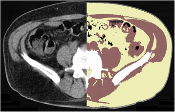
Axial CT image of the abdomen: Left side of the image: original scan/Right side of the image: segmented CT image with staining of the different tissue types (air = black, fat = yellow, muscle = red, and bone = white)

Axial CT image of the abdomen: Left side of the image: original scan/Right side of the image: segmented CT image with staining of the different tissue types (air = black, fat = yellow, muscle = red, and bone = white)