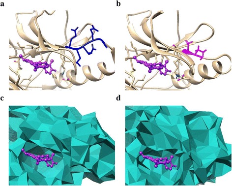
Fig. 1.

a and b show the comparison of the crystal structures of WT EGFR and the mutant delE746_A750insAP. c and d are the alpha shape models of the drug binding pocket of (a) and (b), respectively. The original site is colored blue while the corresponding mutant site is shown in magenta. The drug molecule (gefitinib) is displayed in purple
