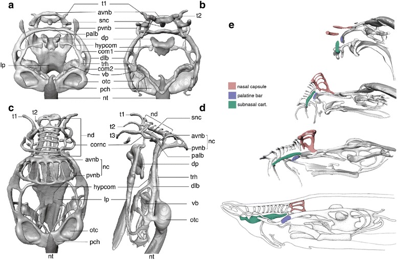
Fig. 2.

Cranial skeletons of the Eptatretus embryo. a Eptatretus burgeri embryo at stage 53 in dorsal view. b E. burgeri embryo at stage 53 in ventral view. c E. burgeri embryo at stage 60 in dorsal view. d E. burgeri embryo at stage 60 in left lateral view. e Relative growth of the developing hagfish chondrocrania. From top to bottom, stage 53, stage 60, prehatching and adult hagfish crania. Note, due to the rostral growth of the snout by addition of nasal duct cartilage and extension of subnasal cartilage (green), that the position of the nasal capsule (red) shifts relatively caudal through development. The palatine bar, or the transverse commissure on the rostral tip of the dorsal longitudinal bar, is colored blue for the reference. avnb, anterior vertical nasal bar; com1, 2, commissures of dlb; cornc, cornual cartilage; dlb, dorsal longitudinal bar; dp, dental plate; hypcom, hypophyseal commissure; lp, lingual plate; nc, nasal capsule; nd, nasal duct cartilage; nt, notochord; otc, otic capsule; palb, palatine bar; pch, parachordal; pvnb, posterior vertical nasal bar; rp, rostral plate; snc, subnasal cartilage; t1-3, cartilaginous support for tentacles; trh, trabecula of hagfish; vb, velar bar
