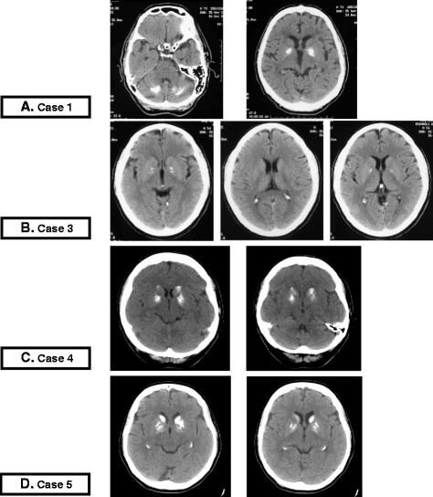
Fig. 1.

CT scan imaging of Cases 1,3,4 and 5. a Case 1. CT scans: extensive bilateral calcification of pallidus nuclei of basal ganglia and dentate nuclei of cerebellum; Hypodense malacic aspect in left frontal lobe. b Case 3. CT scans: mineral hyperdense deposits in bilateral pallidus nuclei without other area of altered density. Small enlargement, age-related, of frontal cerebral sulci due to atrophy. c Case 4. CT scans: hyperdense simmetric calcific deposits in pallidus nuclei and caudate nuclei of both sides. d Case 5. CT scans: hyperdense bilateral calcific deposits in the head of caudate nuclei, putamen and pallidus nuclei
