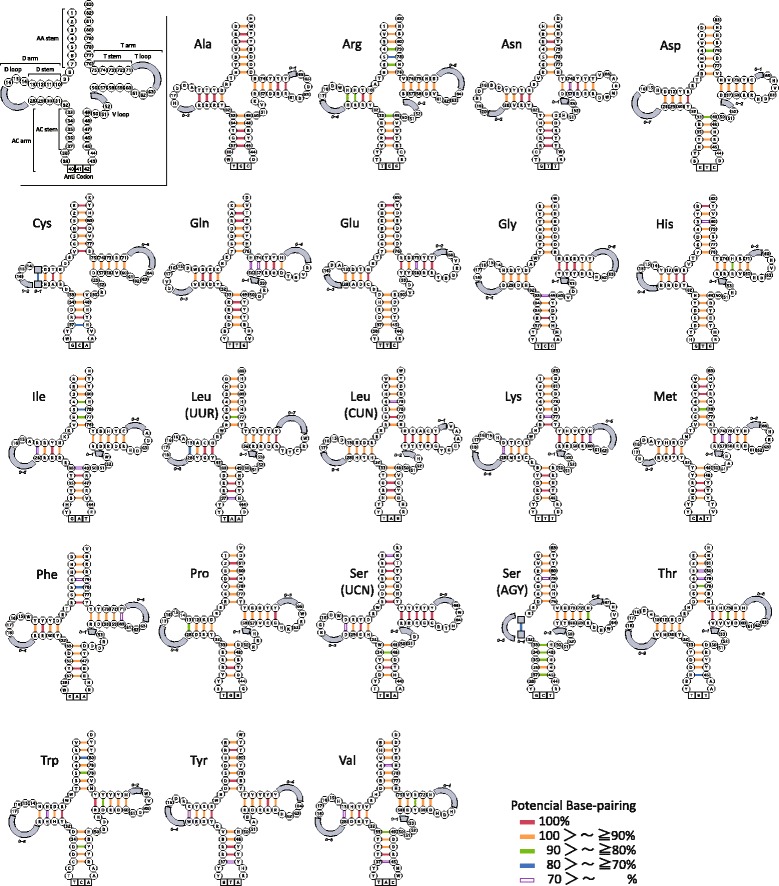
Fig. 4.

Secondary structure model of 22 mitochondrial transfer RNAs (tRNAs) displaying variation in the 250 fishes. In the inset, nucleotide positions common to all tRNAs are numbered from 1 to 83. Stem and loop regions with length variation are denoted by a shaded square and arc, respectively. Conserved nucleotides are shown in the figure by IUB code [89] as follows: K (G, T), M (A, C), N (A, C, G, T), R (A, G), S (G, C), W (A, T), and Y (C, T). Frequencies of Watson–Crick and wobble base pairing observed in the 250 fishes are shown with color-coded bars (see Additional files 8 and 9 for detailed information)
