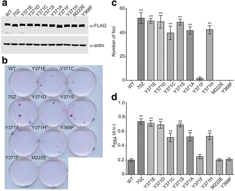
Fig. 6.

Transformation potential of Cbl variants in focus formation assays. a Immunoblot of lysates from 3T3 fibroblasts infected with FLAG-tagged Cbl variants using α-FLAG antibody (top) and α-actin (bottom) antibody as a loading control. b Sulforhodamine B-stained 3T3 fibroblasts infected with indicated Cbl variants. c Mean number of foci formed by Cbl variant-infected 3T3 fibroblasts shown in a bar graph (n = 3). No foci were present in 3T3 cells infected with wild-type Cbl, Cbl M222E or Cbl Y368F. Double asterisks (**) denote significant differences (P < 0.05) between indicated Cbl variant and wild-type using ANOVA followed by Dunnett’s multiple comparisons test. Error bars indicate standard deviation. d As in (c) but for A564 of extracted sulforhodamine B from Cbl-infected 3T3 fibroblasts
