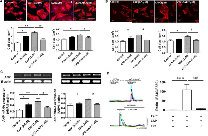
Figure 1.

Activation of TRPV1 induced a cardiohypertrophic response and elevated intracellular calcium level in cultured cardiomyocytes. A, Histological staining of H9C2 cells treated with vehicle, CAP, and CPZ plus CAP for 48 hours is shown; cardiomyocyte cross‐sectional area was measured after treatment with TRPV1 agonist CAP or ANA (6 independent experiments per group, 20 cells counted per experiment). *P<0.05, **P<0.01 versus control, # P<0.05 versus 2 μmol/L ANA, ## P<0.01 versus 2 μmol/L CAP. B, Morphologies of isolated rat neonatal cardiomyocytes were examined after CAP or CPZ plus CAP treatment for 48 hours (5 independent experiments per group, 20 cells counted per experiment), and cardiomyocyte cross‐sectional area was measured after CAP or ANA treatment. *P<0.05 versus control, # P<0.05 versus 2 μmol/L CAP or 2 μmol/L ANA. C, Representative mRNA products of atrial natriuretic peptide in the CAP‐ or ANA‐treated H9C2 cells (6 independent experiments per group) and expression of ANP relative to β‐actin mRNA were assayed. *P<0.05, **P<0.01 versus control, # P<0.05 versus 2 μmol/L CAP or 2 μmol/L ANA group. D, Representative recording of change in intracellular Ca2+ induced by 2 μmol/L CAP with 0 or 2.5 mmol/L calcium in bath solution was reversed by 2 μmol/L CPZ, and the F340/F380 Fura‐2 fluorescence ratio was measured (8 independent experiments per group, 50 cells counted per experiment).***P<0.001 versus calcium‐free bath solution group, ### P<0.001 versus CAP with calcium‐treated group. ANA indicates anandamide; CAP, capsaicin; CPZ, capsazepine.
