
Figure 1.
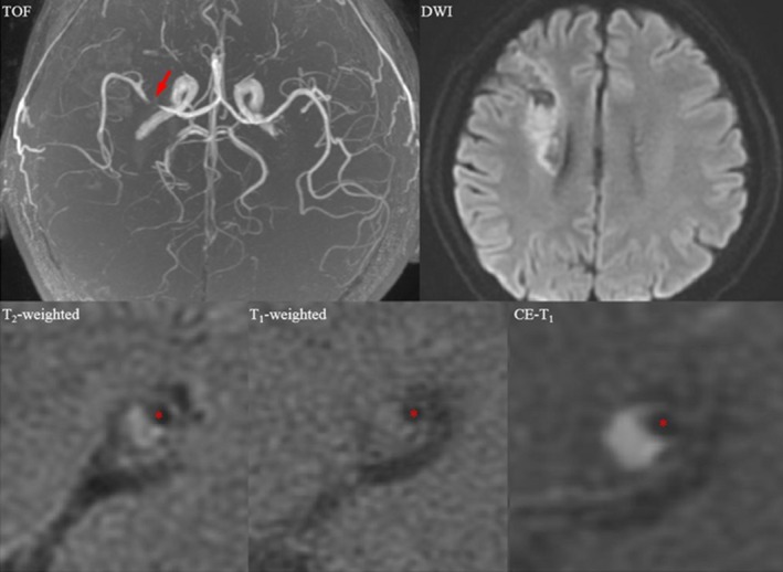
Magnetic resonance images showing an enhancing atherosclerotic plaque in the right middle cerebral artery in a symptomatic patient who had suffered a recent right cerebral hemispheric acute ischemic stroke. A mixture of acute and chronic infarction involving the right periventricular and frontal subcortical regions is seen in the diffusion‐weighted image (DWI); the plaque is shown by an arrow in the time‐of‐flight (TOF) image, and a cross section at the most stenotic site is shown in T2, T1, and contrast‐enhanced (CE) T1 images (red asterisks: lumen).