
Figure 1.
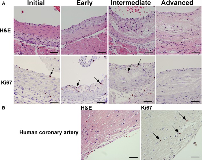
Detection of proliferating cells during murine and human atherosclerotic lesion growth. Ki67 immunohistochemistry and H&E staining in Apoe −/− mice on chow diet identified proliferating cells during various stages of lesion progression, ranging from initial macrophage infiltration to advanced lesions (A). In the initial and early stage, macrophages below the endothelium, as well as foam cells in fatty streaks, were Ki67 positive (Initial and Early, arrows). At 24 weeks of age, in lesions of the intermediate type with large necrotic cores, few Ki67‐positive cells were observed (Intermediate, arrows). At 55 weeks of age, lesions were mostly necrotic with very few Ki67‐positive cells (Advanced). Human lesions from coronary arteries stained with Ki67 contained some Ki67‐positive cells in areas rich in macrophages (B, arrows). Bar=50 μm. H&E indicates hematoxylin and eosin.