Figure 2.

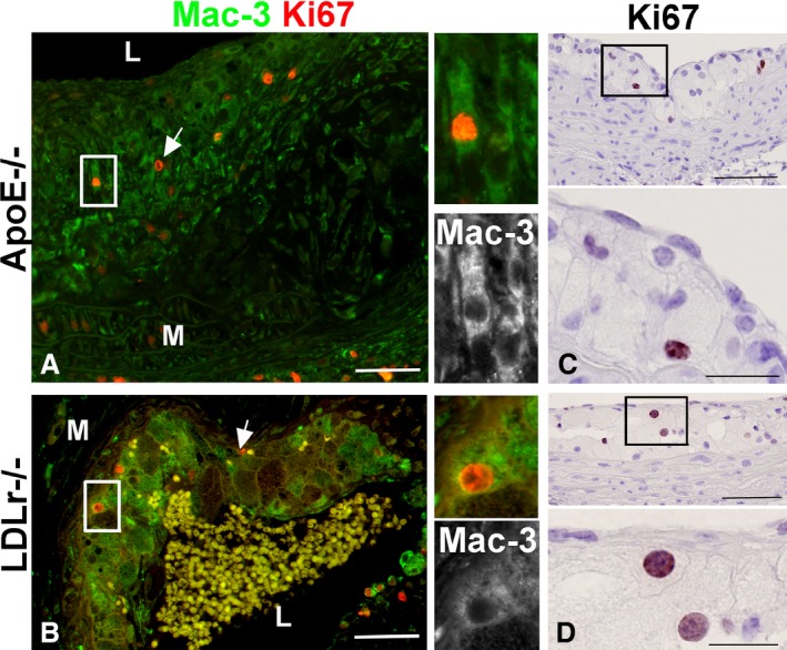
Mac‐3 and Ki67 immunofluorescence of ApoE −/− and LDLR −/− atherosclerotic lesions detects proliferating macrophages. Double immunofluorescence for the macrophage marker Mac‐3 (green) and Ki67 (red) reveals that some of the proliferating cells in the Apoe −/− (A) and LDLr −/− (B) lesions were macrophages. By immunohistochemistry, Ki67‐positive cells exhibited foam cell morphology with large nuclei and lipid vesicles visible at high magnification (insets), in fatty streaks from Apoe −/− mice (C), and from LDLr −/− mice (D). L, lumen; M, media; bar=50 μm, and 20 μm in high‐magnification insets.
