Figure 3.

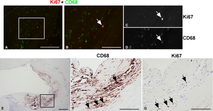
CD68‐ and Ki67‐positive macrophages in human coronary artery atherosclerotic lesions. CD68 staining identified proliferating cells (Ki67 positive) in human coronary arteries as macrophages. Double immunofluorescence of Ki67 and CD68 (A through D), a double‐labeled cell (Ki67, red; CD68, green), was identified in (A) and magnified in (B). The same cell is shown in separate monochrome images (C and D). Lesion areas rich in macrophages were identified by CD68 immunohistochemistry (E) and then analyzed at higher magnification (F). Adjacent sections were stained with Ki67 (G) and showed that Ki67‐positive cells were found in areas rich in macrophages (arrows). Bar=100 μm.
