
Figure 2.
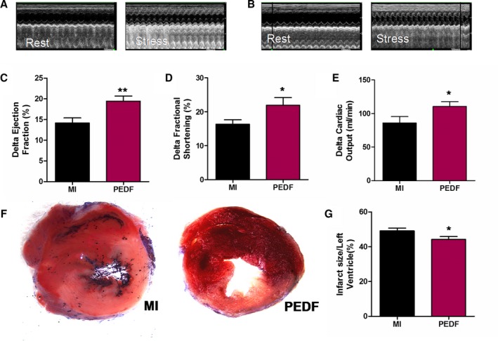
Evaluation of cardiac functional reserve after acute myocardial infarction (AMI). Cardiac function was measured in pigment epithelium‐derived factor (PEDF)‐transfected myocardial infarction (MI) hearts using transthoracic M‐mode echocardiography. Measurements were performed in rats before injection of dobutamine (1 μg/g) and 10 minutes after injection at 2 weeks post‐AMI. The 2 groups included vector transfected rats (MI) and PEDF‐transfected rats (PEDF). A, Representative echocardiograms of an MI rat before and after dobutamine injection. B, Representative echocardiograms of a PEDF‐transfected rat before and after dobutamine injection. C, Delta left ventricular ejection fraction. D, Delta left ventricular fractional shortening. E, Delta left ventricular cardiac output. F, Representative images of 2,3,5‐triphenyltetrazolium‐stained myocardial tissues. G, Quantification of infarct size. Data are shown as mean±SE from 7 hearts (n=7). *P<0.05 versus MI; **P<0.01 versus MI. Results show that PEDF significantly improved cardiac functional reserve and reduced myocardial infarct size when compared to the MI group.