
Figure 5.
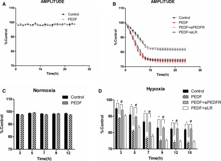
Tracking of contraction changes in neonatal cardiomyocytes. Cardiomyocytes with or without PEDF‐R‐RNAi‐LV or LR‐RNAi‐LV pretransfected were cultured for 24 hours under hypoxia condition. A video edge detection system was used to monitor single‐cell contraction changes. The 4 groups included hypoxia (control), PEDF under hypoxia (PEDF), PEDF with PEDF‐R‐RNAi‐LV under hypoxia (PEDF+siPEDF‐R), and PEDF with LR‐RNAi‐LV under hypoxia (PEDF+siLR). A and C, Amplitude of contraction under normoxia condition. Data are shown as mean±SE from 6 cardiomyocytes (n=6). No significant difference was observed between PEDF and control groups. B, Amplitude of contraction under hypoxia condition. Data are shown as mean±SE from 8 cardiomyocytes (n=8). P<0.001 PEDF versus control; P<0.001 PEDF+siPEDF‐R versus PEDF. D, Statistical analysis of several time points under hypoxia condition. Data are shown as mean±SE from 8 cardiomyocytes (n=8). *P<0.01; # P<0.01. Data are expressed relative to control values measured at the start of each experiment. Results show that PEDF (10 nmol)‐pretreated cardiomyocytes showed a more‐rapid time‐dependent contraction reduction, and the effects could be attenuated by PEDF‐R interference. No significant difference was observed between PEDF and PEDF+siLR groups. LV indicates lentivirus; PEDF, pigment epithelium‐derived factor; PEDF‐R, PEDF receptor.