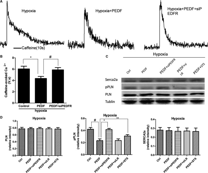
Figure 7.

Sarcoplasmic reticulum (SR) Ca2+ load and proteins expression in neonatal cardiomyocytes. Another group added as PEDF (10 nmol) with staurosporine (STS) under hypoxia (PEDF+STS). A, Representative caffeine‐induced Ca2+ transients from Hypoxia, Hypoxia+PEDF, and Hypoxia+PEDF+siPEDF‐R neonatal cardiomyocytes. B, Quantification of the area under caffeine‐evoked Ca2+ transients in rat ventricular myocytes. Data are shown as mean±SE from 6 cardiomyocytes (n=6). *P<0.05; # P<0.05. Results show that PEDF reduced sarcoplasmic reticulum (SR) Ca2+ load and PEDF‐R interference also could attenuate the effect. C and D, Sarcoplasmic/endoplasmic reticulum Ca2+‐ATPase (Serca2a), phosphoylated phospholamban (pPLN), and phospholamban (PLN) expression in hypoxic cardiomyocytes. Data are shown as mean±SE from 3 samples (n=3). *P<0.01; # P<0.01; **P<0.05. Results show that PEDF reduced PLN phosphorylation under hypoxia condition through PEDF‐R and the protein kinase C alpha inhibitor, staurosporine, could attenuate the effects of PLN dephosphorylation. No significant difference was observed in Serca2a and PLN. PEDF indicates pigment epithelium‐derived factor; PEDF‐R, PEDF receptor.
