
Figure 8.
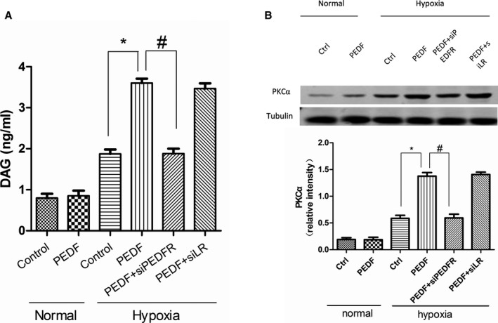
Diacylglycerol (DAG) content and protein kinase C alpha (PKCα) regulation in neonatal cardiomyocytes. ELISA analysis were used to test DAG content. A, Quantification of DAG content in cardiomyocytes after 12 hours of hypoxia. Data are shown as mean±SE from 4 samples (n=4). *P<0.001; # P<0.001. Results show that PEDF (10 nmol) induced diglyceride increase in hypoxic cardiomyocytes through PEDF‐R and had no effects under normal condition. B, PKCα protein expression in cardiomyocytes. Data are shown as mean±SE from 3 samples (n=3). *P<0.01; # P<0.01. Results show that PKCα significantly increased in the PEDF group compared to the control group in hypoxic cardiomyocytes. PEDF indicates pigment epithelium‐derived factor; PEDF‐R, PEDF receptor.