Figure 6.

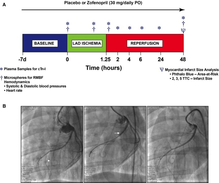
Experimental protocol for in vivo myocardial ischemia/reperfusion (I/R) injury in swine. A, Female Yucatan pigs were subjected to 75 minutes (1.25 hours) of myocardial ischemia by occluding left anterior descending coronary artery (LAD) via a balloon catheter placement and 48 hours of reperfusion. Zofenopril (30 mg/daily PO) or placebo therapy was initiated at 7 days prior I/R injury and continued for 2 days after ischemia. At baseline, 60 minutes of LAD occlusion, 15 minutes of reperfusion, and 48 hours of reperfusion, microspheres labeled with samarium, europium, lutetium, or lanthanum were injected to measure regional myocardial blood flow (RMBF). At baseline, 60 minutes of ischemia, and 15 minutes, 2, 4, 6, 24, and 48 hours of reperfusion, plasma samples were collected for measurement of cardiac troponin‐I release. At day 2 of reperfusion, heart tissue was collected for infarct size determination. Baseline plasma samples were used for assessment of circulating levels of H2S, sulfane sulfur, NO 2 −, and S‐nitrosothiols (RXNO). B, Angiographic left anterior oblique caudal images of the LAD at baseline (left), during 75 minutes occlusion (middle), and at 15 minutes reperfusion (right). Intracoronary occlusion was achieved by inflation of an angioplasty balloon catheter (arrow) deployed in the proximal LAD distal to the first anterior septal branch. Proximal LAD deployment of the balloon produced an area‐at‐risk (AAR) region ≈45% of the left ventricular (LV) mass. The AAR was distributed primarily in the anterior LV free wall and includes a portion of the anterior septum. For measurement of regional blood flow, microspheres were injected into the LV cavity through a pigtail catheter (arrowhead).
