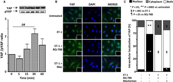
Figure 9.

YAP activation and intracellular localization of YAP after exposure of HK‐2 cells to ET‐1. A, Time course of YAP activation, expressed as the ratio of YAP to phosphorylated YAP, showed YAP activation 30 minutes after ET‐1 exposure. Graph bars represent mean values ± SEM. B, Immunofluorescence for YAP (green) in HK‐2 cells after exposure to ET‐1, in the presence or absence of either macitentan or BQ‐788. In untreated HK‐2 cells the immunosignal was clearly evident in both nucleus and cytoplasm. ET‐1 induced blunting of the YAP signal in the cytoplasm and its increase in the nucleus, indicating dephosphorylation of YAP and translocation to the nucleus, where it can trigger transcription of genes involved in EMT. Pretreatment with either macitentan or BQ‐788 prevented the ET‐1–induced increase of YAP immunosignal in the nucleus. Nuclei were labeled with DAPI (blue); the omission of primary antibody confirmed the specificity of the reaction (insets). C, Intracellular YAP localization. Quantitative analysis confirmed that ET‐1 favors translocation of YAP to the nucleus (vs untreated cells) and that macitentan and BQ‐788 prevented such translocation. For all experiments mean values of 3 independent experiments, each in duplicate, are reported.
