Figure.
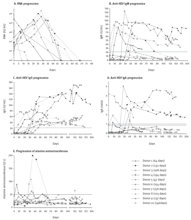
Progression of hepatitis E virus RNA, IgM, IgG and IgA antibodies and alanine aminotransferase in blood donors with autochthonous hepatitis E virus genotype 3 infection, Germany, 2011 (n = 10)
The day of the detection of HEV RNA by PCR screening was defined as day 0. Grey-shaded areas (panels B–D): cut-off values of the different serological assays as described in the Methods section. Solid horizontal line (panel E): reference range of 0–50 U/L. Days in brackets after donor legend: time period were samples were taken. Ratio: extinction sample/calibrator.