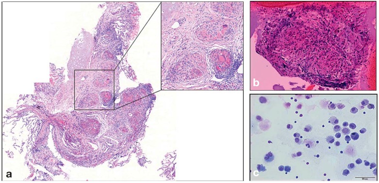
Figure 3.
Cytological and histological findings in sarcoidosis
a) transbronchial lung biopsy specimen, with non-caseating granulomas containing epthelioid and giant cells
b) a granuloma obtained by ultrasound-guided needle biopsy of a mediastinal lymph node
c) cell smear of a bronchoalveolar lavage (BAL) specimen, showing the typical abundance of CD4+ T lymphocytes (small, round cells with a large nucleus and little cytoplasm). Most of the other cells are alveolar macrophages with a large amount of cytoplasm.

