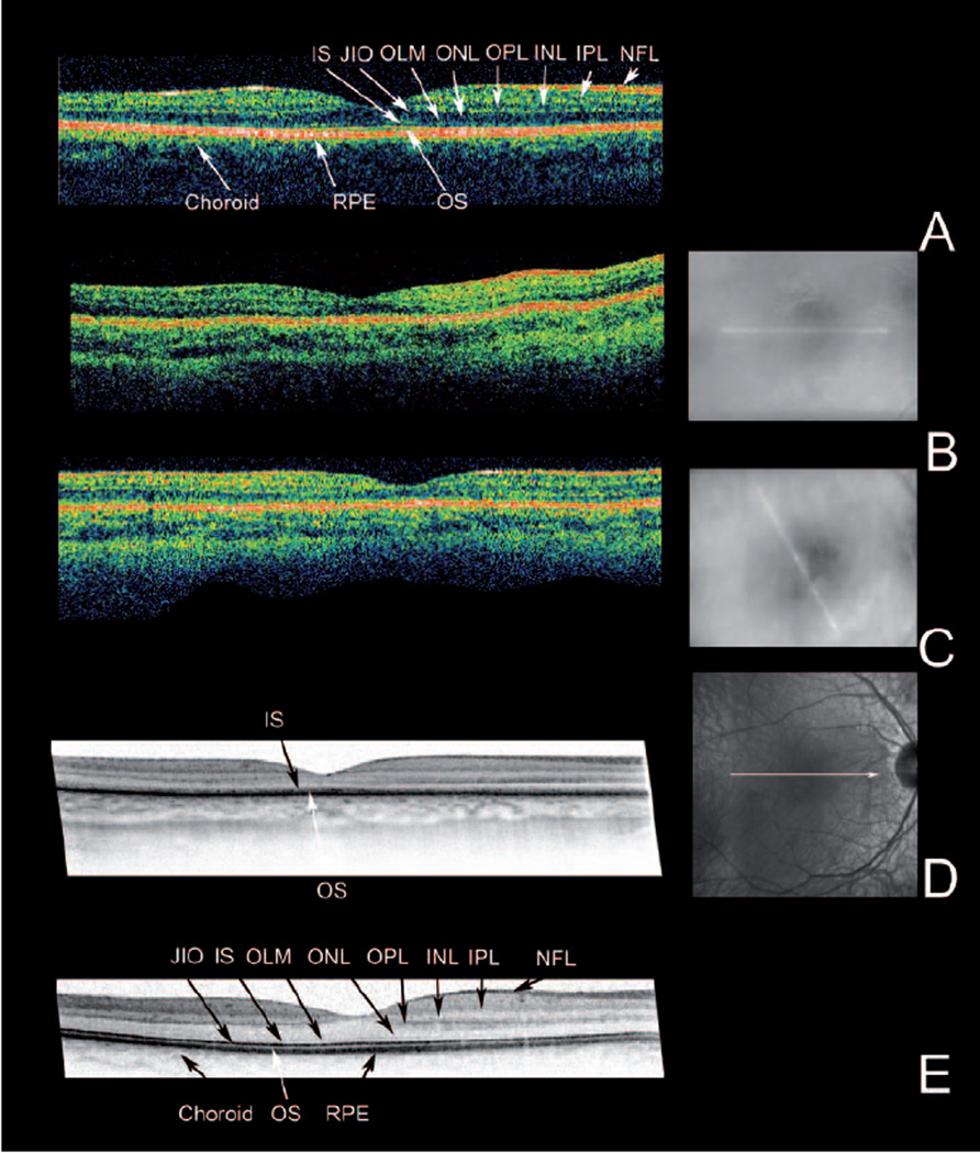
Figure 2.

OCT imaging. For each eye the OCT image is accompanied by a fundus image showing the position of the scan. The findings were similar in both eyes. Only right eyes are shown.
(A) Stratus OCT recording of a 20-y old normal proband. (B) and (C) Stratus OCTs of the patient at age 3 and 7-y. (D) At the age of 7-y, a high resolution OCT was recorded with a Spectralis HRA-OCT. For comparison (E) a Spectralis OCT recording of the 20-y old normal proband is included. Note the overall reduced thickness of the retina in the patient (B, C, D). Spectralis imaging (D) shows that this loss of thickness is due to the loss of stratification in the inner (IS) and outer segment layers (OS) and thinning of the outer nuclear layer (ONL). NFL: Nerve fibre layer, IPL: inner plexiform layer, INL: inner nuclear layer, OPL: outer plexiform layer, ONL: outer nuclear layer, OLM: outer limiting membrane, IS: inner segment layer, JIO: junction between inner and outer segments, OS: outer segment layer: RPE: retinal pigment epithelium
