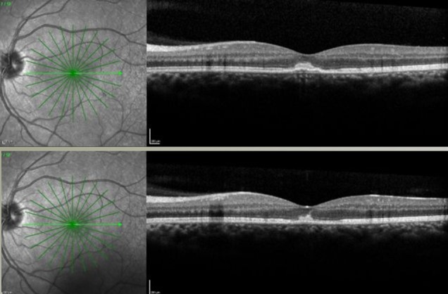
Figure 2. OCT imaging with horizontal section of the left macula 3 days (top) and 6 days (bottom) after the event. Note the hyperreflective foveal lesion, initially prominent, and reduced 3 days later with at that time also regression of the hemorrhage to half size.