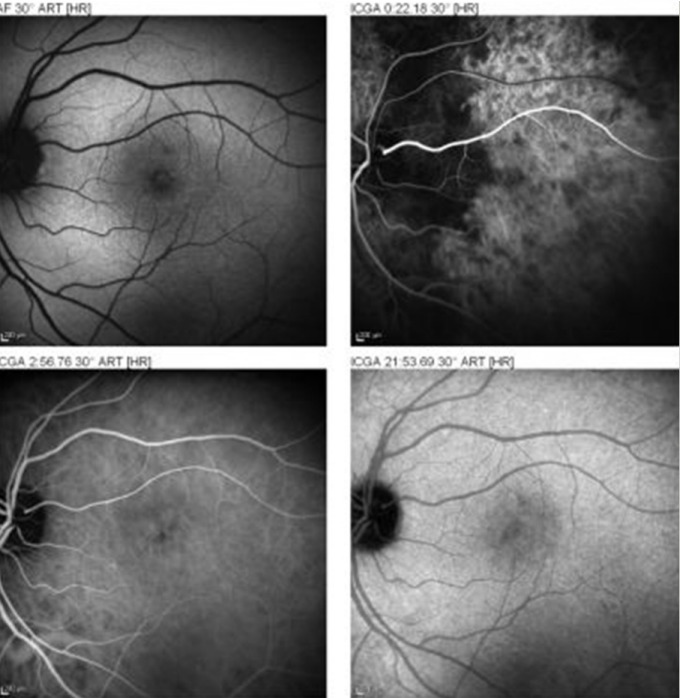
Figure 3. Six days after the event the ICG angiogram shows in the early phase (top right) focal choroidal non perfusion in the papillomacular and macular area, compatible with delayed filling in a watershed zone. In midphase and late phase ICG angiogram (bottom left and right) no anomalies are noted.