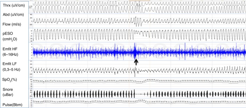
Fig. 2.
Example of a 5-min polysomnography period. At the beginning of the sheet, respiratory movements are stable; flow channel shows slight flow limitation and mouth breathing. Negative esophageal pressure is increased up to −30 cm H2O. Emfit high-frequency channel shows multiple spikes. At the middle of sheet (marked with a black arrow) is a short arousal with opening of upper airway, normalizing esophageal pressure values and cease of spiking. Gradually breathing effort starts to increase again. Channels from top: thoracic and abdominal belts, flow by nasal pressure transducer, esophageal pressure, Emfit high-frequency channel, Emfit low-frequency channel, arterial oxyhemoglobin saturation, snoring, and pulse.

