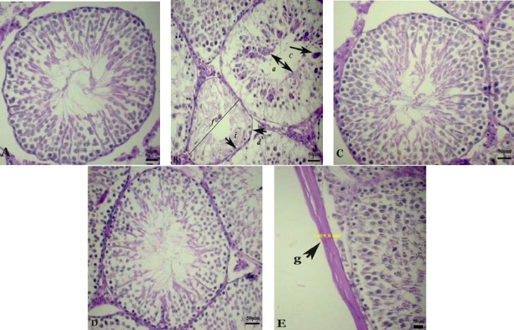
Figure 4.
Photographs of several seminiferous tubules in rats stained with H&E (× 400). Testicular microscopic image of (A) normal rat testis of C group; (B); Diabetic rat testis with disorganization of the seminiferous tubule epithelium (a) and separation of the basement membrane (b), giant multinucleated cell in seminiferous tubule (c), seminiferous tubular atrophy (d), decline of germ cell layers number (h), spermatogenous arrest (i), the diameter of seminiferous tubules (f); (C) normal rat testis that received RJ; (D) diabetic rats treated with RJ showed improvement in seminiferous tubular structure; (E) the thickness of tunica albuginea indicated by arrows (g).

