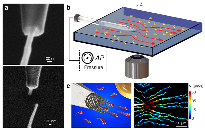
Figure 1. Nanojet experimental set-up.
(a) SEM image of a CNT insertion into a nanocapillary (top) and after sealing (bottom). The CNT has dimensions (Rt, Lt)=(50,1000) nm. (b) Sketch of the fluidic cell used to image the Landau-Squire flow set-up by nanojets emerging from individual nanotubes. (c) (Left) Sketch of a nanotube protruding from a nanocapillary tip. (Right) Trajectories of individual colloidal tracers in a Landau-Squire flow field in the outer reservoir. The flow was driven by a nanojet from a CNT whose dimensions were (Rt, Lt) =(33,900) nm, with ΔP=1.7 bar. Both reservoirs contained water with 10-2 M KCl.

