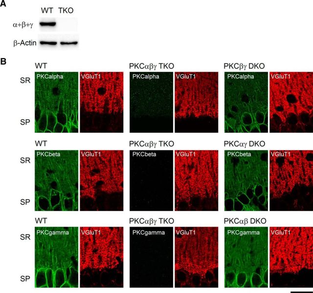
Figure 2.
Expression of PKC isoforms in the hippocampal CA1 region. A, Western blot of classical PKC isoforms from WT and PKCαβγ TKO brain lysates. B, Confocal images of Ca2+-dependent PKC isoforms α, β, and γ in the hippocampal CA1 region from the indicated genotypes. SP, Stratum pyramidale; SR, stratum radiatum. Scale bar, 25 μm.

