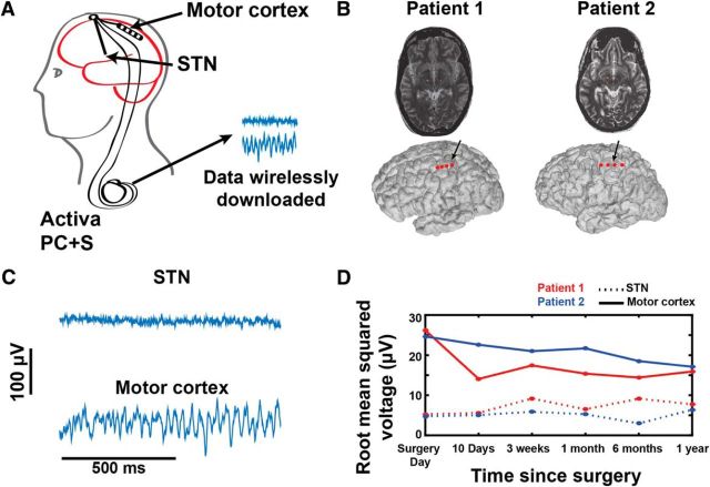
Figure 1.
Electrode locations, raw signals, and signal stability over time. A, Schematic of the Activa PC+S. B, Electrode locations (indicated in red) for Patients 1 and 2 over cortex (top) and in STN (bottom). Locations are derived from the preoperative MRI merged with the intraoperative CT. C, Raw signals from STN and Motor cortex. D, Root mean square voltage off DBS for motor cortex ECoG potentials and STN LFP for each patient, recorded over 12 months. Signals are relatively stable over time.

