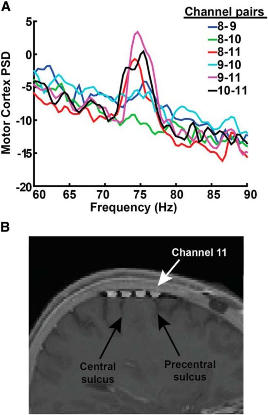
Figure 13.

Optimal recording location for cortical gamma oscillations. A, Set of recordings from all contact pairs recorded sequentially (over a time period of 3 min) from Patient 2 during dyskinesia. This recording was obtained with a sampling rate of 422 Hz (required to obtain a rapid sequential “montage recording” across all possible contact pairs) with DBS off. The peak is most strongly observed in contact pairs that include contact 11. The PSD scale is 10 * log10(μV2/Hz). B, Fusion of intraoperative CT with preoperative MRI, showing contact locations with respect to gyral anatomy. Contact 11 (white arrow) is located over the anterior portion of the precentral gyrus as well as precentral sulcus.
