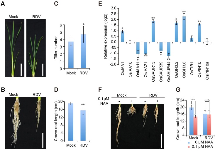
Fig 1. RDV infection disturbs auxin pathway in rice.
(A and B) Aboveground (A) and root (B) phenotypes of mock- and RDV- infected rice plants at 6-week-old seedling stage. Bars: 10 cm. (C and D) Schematic representation of the tiller number and crown roots length of mock- and RDV- infected rice plants in (A) and (B). The average (± standard deviation (SD)) values were obtained from three biological repeats, with 15 plants from each line in every repeat. Significant differences were indicated (*P<0.05, **P<0.01) based on Student’s t-test. (E) Relative average expression (log2) of auxin-induced genes in RDV-infected rice plants. Data were obtained from qPCR assays and analyzed using 2-ΔΔC(t) method and the OsEF1a mRNA levels were used as internal controls. Values are mean ± SD (n = 3 biological replicates). Columns with asterisks are statistically different according to Student’s t-test (*P<0.05, **P<0.01) as compared to their expression in mock-inoculated rice plants. (F and G) RDV-infected rice plants exhibit reduced sensitivity to auxin treatment. Phenotypes (F) and lengths (G) of crown roots of mock- and RDV- infected 4-week-old seedlings cultured in liquid nutrition containing 0 or 0.1 μM NAA for 10 days. Bar: 10 cm. The average (± SD) values were from three biological repeats with 15 plants for each line every repeat. Significant differences were indicated (n.s., no significant, **P<0.01) based on Student’s t-test.

