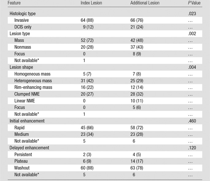
Table 1.
MR Imaging Features of Index Lesions and Dominant Additional Lesions

Note.—Unless otherwise indicated, data are numbers of lesions, and data in parentheses are percentages. NME = nonmass enhancement.
*Not included in the test. No residual enhancement was seen after biopsy of the index lesion, and only the clip was visible at MR imaging.
