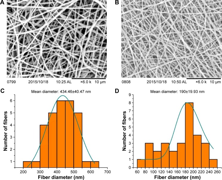
Figure 1.
Nanofiber morphology and diameter distribution of fabricated electrospun membranes.
Notes: Representative SEM images of PU (A) and bio-nanofibrous membrane (B). Diameter distribution histogram of PU (C) and PU-HN-PA (D) dressing materials.
Abbreviations: HN, honey; PA, Carica papaya; PU, polyurethane; SEM, scanning electron microscopy.

