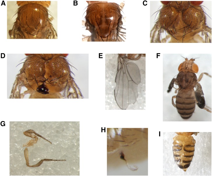
Figure 2.
Examples of dysmorphologies produced by the tubGS driver in the presence of 10 μM RU486. (A–D) Thoracic abnormalities: (A) missing scutellar part, (B) mild cleft, (C) severe cleft, and (D) necrotic tissue, always localized at the scutellum or notum. (E and F) Wing abnormalities: (E) notched wings, with notches localized on the marginal anterior or posterior side or both, (F) noninflated wings. (G) Leg abnormalities, including overgrown, reduced, and fused leg segments, sometimes present all together. (H) Externalized trachea, always in the ventral abdomen. (I) Abdominal clefting: strong midline splits between all dorsal tergite plates; laterotergites do not fuse at the dorsal midline and remain as hemitergites, with incomplete fusion of abdominal epidermis. These phenotypes were seen in all genetic backgrounds tested (OregonR, CantonS, w1118, and wDAH). tubGS; α-tubulin-GeneSwitch.

