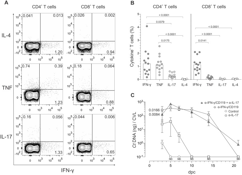
Fig 4. C. trachomatis-specific CD4+ T cells displayed TH1 and TH17 effector function, but control of ivag C. trachomatis challenge is mainly mediated by TH1 immunity.
At 60 dpi, mice were ivag challenged with 106 IFU of C. trachomatis serovar D. At 5 dpc, animals were euthanized, DLN excised and processed into single-cell suspensions, and cells incubated with inactivated Chlamydia EB or media alone. (A) Representative contour plots from the intracellular cytokine staining (ICS) assay used to quantify secretion of IFN-γ, TNF, IL-17, and IL-4 by CD4+ and CD8+ T cells that responded to stimulation with Chlamydia EB; (quadrant numbers indicate percentages of cytokine-producing cells). (B) Percentages of cytokine-producing CD4+ and CD8+ T cells; (n = 13) (bars indicate medians). (C) Using additional Balb/cJ mice at 60 dpi, 106 IFU of C. trachomatis serovar D was ivag administered to untreated controls or mice treated with antibodies blocking IFN-γ, IL-17, or IFN-γ and IL-17 signaling concomitant with challenge. At various days after challenge, CVL specimens were collected to measure Chlamydia DNA levels via RT-qPCR. AUC analysis for bacterial clearance revealed that Chlamydia challenge was primarily controlled by TH1 immunity; (n = 5 per group) (values are means ±SD) (results representative of 2 independently performed studies) (ND, non-detectable).

