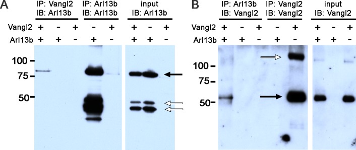
Figure 7.
Arl13b biochemically interacts with Vangl2. (A) Protein extracts from HEK293-T cells transiently transfected with Vangl2 and/or Arl13b and immunoprecipitated (IP) with antibodies against Vangl2 (lanes 1–3) or Arl13b (4–5) and subsequently immunoblotted (IB) with antibodies against Arl13b. Immunoblots of input samples show the presence of Arl13b from different extracts used for IP (lanes 6–8). Black arrows indicate bands corresponding to Arl13b, while open arrows indicate nonspecific bands. (B) Protein extracts from HEK293-T cells transiently transfected with Vangl2 and/or Arl13b and immunoprecipitated with antibodies against Arl13b (lanes 1–3) or Vangl2 (lanes 4–5) and subsequently subjected to IB with antibodies against Vangl2. Immunoblot of input samples to show the presence of Vangl2 in different protein extracts (lanes 6–8, black arrow). The high molecular weight band (white arrow) likely represents Vangl2 homodimers. Molecular mass of protein markers is denoted in kDa.

