Figure 4.
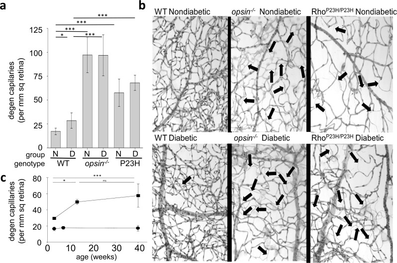
Effect of diabetes and opsin mutations on the retinal vasculature. (a) A graphical summary of the number of degenerate (degen) capillaries in retinas from nondiabetic and diabetic C57Bl/6J (WT), opsin−/−, and RhoP23H/P23H (P23H) mice at 10 months of age (8 months of diabetes; n = 5–14 in all groups). (b) Representative photomicrographs demonstrating the capillary degeneration at the same age. Degenerated retinal capillaries (acellular capillaries; illustrated by black arrows) are significantly more numerous in WT diabetic mice compared to WT nondiabetic mice, but the capillary degeneration is substantially exacerbated in nondiabetic or diabetic mice deficient in opsin or expressing the P23H mutant opsin. Unlike what was seen in WT mice, diabetes did not exacerbate retinal capillary degeneration in opsin−/− and P23H mice compared to their appropriate nondiabetic controls. (c) Degeneration of retinal capillaries is accelerated during the first 3 months of life (while photoreceptors are present but degenerating) in nondiabetic P23H mice (squares) compared to nondiabetic WT controls (circles). Vascular degeneration slows after photoreceptors have degenerated (after 3–4 months); n = 3 to 14 in all groups. Vascular histopathology was quantitated microscopically following isolation of the vasculature by the elastase digestion method. Horizontal lines indicate statistically significant differences between groups; absence of a bar indicates no statistically significant difference. N, nondiabetic; D, diabetic. *P < 0.05; ***P < 0.001. Mean ± SD.

