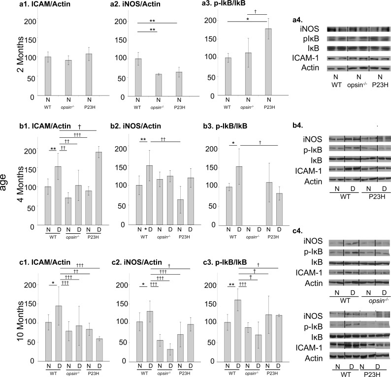
Figure 7.
Effect of opsin deletion and the knock-in of P23H mutant opsin on retinal expression of proinflammatory proteins in mice. Data summarize expression of ICAM, iNOS, and the ratio of phosphorylated IĸBα to total IĸBα at 2 (a1–4), 4 (b1–4), and 10 (c1–4) months of age (0, 2, and 8 months of diabetes). Data at 2 months of age do not include diabetic animals because diabetes was not induced until 2 months of age. Data used to calculate expression of ICAM1 and iNOS by the retinas of opsin−/− mice nondiabetic and diabetic for 2 months (4 months of age) were published previously28 but are regraphed here to show the expression patterns relative to the WT nondiabetic C57Bl/6J group. Figures a4, b4, and c4 show representative immunoblots. Areas where the membranes were cut to remove omitted lanes are indicated by vertical lines. Inflammatory proteins were quantitated by immunoblots of retinal homogenates, and expressed relative to actin in the same sample. n = 3 to 7 in all groups. *P < 0.05 or **P < 0.01 or ***P < 0.001 compared to N C57Bl/6J controls; †P < 0.05 or ††P < 0.01 or †††P < 0.001 compared to D C57Bl/6J controls. N, nondiabetic; D, diabetic.

