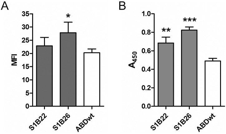
Fig 8. Flow cytometric (A) and whole-cell ELISA (B) analyses of binding of recombinant Stx1B by L. lactis cells displaying S1B variants or ABDwt on their surface.
(A) Alexa488-conjugated Stx1B was used for detection. MFI: Mean fluorescence intensity. (B) Mouse antiStx1B antibody and HRP-conjugated anti mouse antibody were used for detection of Stx1B. A450: Absorbance at 450 nm. Vertical bars denote standard deviation. MFI or A450 values of S1B binders were compared to those of the ABDwt control using Student’s t test. *: p<0.05, ** p<0.01, *** p<0.001.

