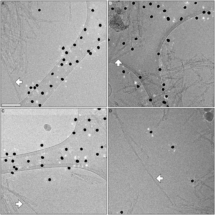
Fig 4. Additional cryo-EM micrographs of GPI-anchorless prion fibrils.
Electron micrographs showing representative GPI-anchorless prion fibrils, including four isolated fibrils that were subsequently analyzed by image processing (white arrows). The labels (A to D) correspond to the lettering in the 3D fibril reconstruction figures (vide infra). Black dots originate from fiducial gold that was added for tomographic studies. Scale bar, 100 nm.

