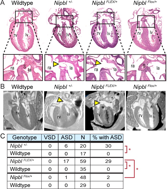
Fig 3. Nipbl FLEX/+ and Nipbl+/- mice develop heart defects at the same high frequency.
A, B. Paraffin-sectioned hearts stained with H&E (A) and MRI-scanned hearts (B) show large atrial septal defects (yellow arrowheads) in Nipbl+/- and NipblFLEX/+ mice, but not in wildtype or NipblFlox/+ mice. Scans and histology were performed on fixed tissue from E17.5 embryos. Scale bar = 500 μm. la, left atrium; lv left ventricle; ra, right atrium; rv right ventricle; S, septum. C. Summary table showing incidence of atrial septal defects (ASDs) and ventricular septal defects (VSDs) in hearts of Nipbl+/-, NipblFLEX/+, NipblFlox/+ mice and wildtype littermate embryos at E17.5. Asterisks: p < 0.01 by Chi-square analysis. Data were pooled from analyses of multiple crosses (see S1 Data: Sample Numbers) and progeny are on various backgrounds depending on parental backgrounds: Nipbl+/-, CD-1; NipblFLEX/+, mixed; NipblFlox/+, C57Bl6/J or mixed.

