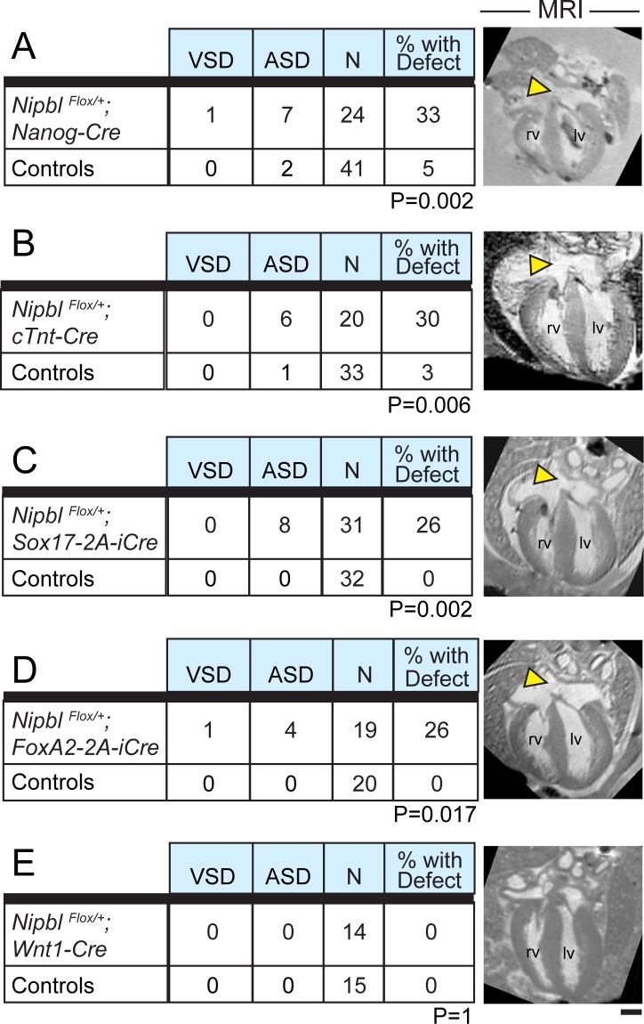
Fig 5. Creation of Nipbl deficiency in cardiac developmental lineages.
NipblFlox/+ mice were crossed with mice hemizygous for each of five indicated Cre-expressing transgenes. MRI analysis of hearts was performed at E17.5. A–D show that whether Nipbl was made deficient in all tissues (Nanog-Cre, A), specifically in cardiomyocytes (cTnt-Cre, B), or primarily in endoderm-derived tissues (Sox17-2A-iCre, C) or mixed cardiac lineages (FoxA2-2A-iCre, D), the incidence of CHDs (primarily ASDs) was approximately 30%. (Chi-square analyses indicate that frequencies of heart defects observed in embryos with Nipbl deficiency in experiments A–D do not differ significantly from each other [p > 0.4 for each pairwise comparison].) In contrast, control hearts (a mix of wildtype, NipblFlox/+, and Cre+ littermates for each specific cross; see S1 Data: Sample Numbers) had an incidence of heart defects ranging from 0%–5% (A–D). Nipbl deficiency in the Wnt1 domain (neural crest) did not give rise to heart defects (E). CHDs occurred primarily in the form of ASDs of the ostium secundum type (yellow arrowheads); VSDs observed in A and D are of the perimembranous type. p-Values are from Chi-square analyses and are indicated for each corresponding cross. Scale bar = 500 μm. lv, left ventricle; rv, right ventricle.

