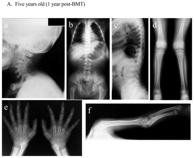
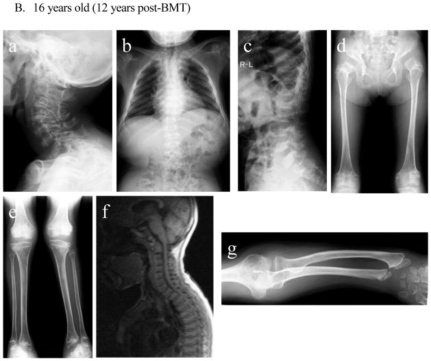
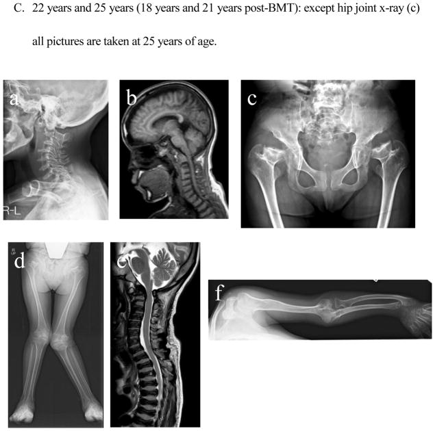
Figure 3. X-ray and MRI photographs after BMT at five years, 16 years, 25 years (Case 1: 1 year, 12 years, and 21 years post-BMT).
- Cervical spine: Dysmorphic changes (platyspondyly and oval shape with anterior beaking) of the cervical vertebra with widening of disc spaces are observed. The thickness of the cervical spine itself is reduced in order from the second cervical vertebra, and intervertebral distance is narrowed. No evidence of cervical spine instability is seen.
- Chest and hip: The upper thorax is narrow. The thorax is also squeezed to the left diaphragm elevated. Formation of the acetabular bottom is wrong. The femoral head is recessed in the femoral neck that has been reduced itself. Both femoral heads with dysmorphic changes are in a position of subluxation.
- Spine: Thoracolumbar body is thin and flat with marked osteodysplasia. Tongue-like anterior beaking of the lumbar vertebrae appears. The vertebrae spaces are wide.
- Lower extremities: Genu valgum is observed. Expansion of both distal end of the femur and the proximal end of the tibia are seen.
- Hands: Hand phalanges, metacarpals, and carpal have marked dysplasia. Bilateral hand radiographs show the cone-shaped tapering of the distal portion of metacarpals 2 through 5, expansion of the radius distal end, and shortening of the ulna distal end.
- Upper extremity: Both proximal and distal ends of the humerus and the forearm bones are swollen, and the forearm bones are curved. The hands take on a characteristic with tilting of the radial epiphysis towards the ulna, resulting from a combination of metaphyseal deformities, hypoplasia of the bones, and degradation of connective tissues.
- Cervical spine: Compared to that at 5 years old, ossification failure of the entire cervical spine has become more sophisticated. Dysmorphic changes (platyspondyly and oval shape with anterior beaking) of the cervical vertebra with widening of disc spaces are not changed.
- Chest: Oar shaped ribs remain wide and are over constricted in their paravertebral portions.
- Spine: Bony changes of the lumbar spine with anterior inferior beaking and platyspondyly of the vertebrae remain evident.
- Hip and Femur: Acetabular space is more expanded in a position of subluxation, and the femoral cap is not observed. There are an oblique acetabular roof with coxa valgus deformity and flared iliac wings. Coxa valga appears, and genu valgum is improving.
- Knee and tibia: Persistent genu valgum is seen (left leg is more severe than right leg). The medial tilt of the ankle mortise is noted.
- There is no evidence of cervical myelopathy due to compression of cervical spinal cord in the first and second cervical vertebrae. Platyspondyly of cervical vertebrae is observed.
- The tapered irregular distal radius and ulna are observed with tilting of the radial epiphysis towards the ulna. Remarkable swelling of the humeral and radial distal end is prominent. Ossification failure of carpal is notable.
- Cervical spine: Dysmorphic changes of the cervical vertebra with widening of disc spaces remain present. Cervical vertebrae become thinner, but the extent of ossification is kept. No evidence of cervical spine instability is seen.
- Cervical spinal cord: Mild compression of the spinal cord is seen, but cervical myelopathy is not noticed.
- The femoral neck has been damaged and shortened. Ossification of the entire pelvis is well maintained. Oblique acetabular roof with coxa valgus deformity and flared iliac wings are observed. Subluxation of the hip joints is seen.
- Bilateral genu valgus can be seen, and intense deformation and swelling of the knee joint are observed. The mild curvature of the femur can be seen.
- MRI of spine shows slight compression of the cervical spinal cord. Universal platyspondyly with anterior beaking is observed in cervical and thoracic vertebrae.
- An elbow joint is swollen, and its destruction is in marked. The curvature of the ulna and radius is seen with tilting of the radial epiphysis towards the ulna in the distal end. Cortical thinning and mild widening of the diaphysis of the humerus are visible.


