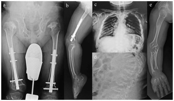
Hip: Femoral head is fitted into the femoral neck, and femoral neck is rudimentary. The acetabular roof is destroyed, and the iliac has been scraped off the top with flared iliac wings. Coxa valga is seen. Both femoral heads are well positioned.
Knee and lower extremity: Knee joint is hypoplasia of the bone, defined as severe. Femur and the entire lower leg have ossification failure.
Chest: Thorax diaphragm is lifted, and chest cavity is compressed. The space between the ribs is also narrow, and the deformation of the ribs is seen, leading to a reduction in air content of the thoracic cavity inferred.
Thoracolumbar spine: Thoracolumbar vertebral bodies are flattening, and anteriorly beaking like tongue-shaped deformation. The intervertebral body space is enlarged. Platyspondyly and anterior beaking of thoracolumbar vertebra were almost unchanged 11 years later post-BMT.
Upper extremity: Due to a high degree of dwarfism, humerus and forearm bone are shortened, and swelling of the elbow is marked. Destruction of elbow joint leads to a strong contracture and the extension limit. Shortening of the metacarpal bone and carpal ossification of failure are marked.

