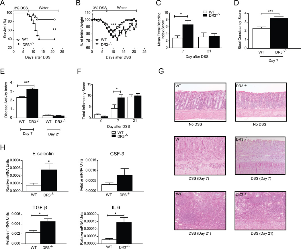
FIGURE 1. Increased severity of acute DSS colitis in DR3−/− mice.
DSS (3% w/v) was added to the drinking water of DR3−/− (n=25) and WT DR3+/+ (n=20) mice for 7 days. A, Increased mortality, and B, increased loss of body weight in DR3−/− compared to WT littermates. C-F, Significant differences between DR3−/− and WT mice were observed by day 7 post-DSS administration, but not day 21, in mean fecal bleeding index scores (C), stool consistency scores (D), disease activity index scores (E), and total inflammatory score (F), calculated by adding the values for individual semi-quantitative histological scoring indices for % ulceration, % re-epithelialization, and active, chronic, and transmural inflammation. G, Representative H&E-stained sections depict the histologic characteristics of colonic tissues from DR3−/− and WT mice, with and without induction of DSS-colitis. DR3−/− mice at day 7 show almost complete loss of the epithelium, with increased infiltration of inflammatory cells in the lamina propria (left middle panel); by comparison WT DR3+/+ mice at day 7 still maintain cryptal architecture with slightly decreased inflammatory cells in laminar propria (right middle panel). DR3−/− and WT mice at day 21 show comparable severity of colitis (left and right lower panels).
Original magnification of photomicrographs, 20x. H, Total RNA was extracted from colonic specimens of DSS-treated DR3−/− and WT DR3+/+ mice. Mucosal mRNA expression of the indicated factors was quantified by real-time PCR. The relative expression of each target gene was normalized to the relative expression of GAPDH in the same sample. All data presented as mean ± SEM; *P < 0.05, **P < 0.01, ***P < 0.001.

